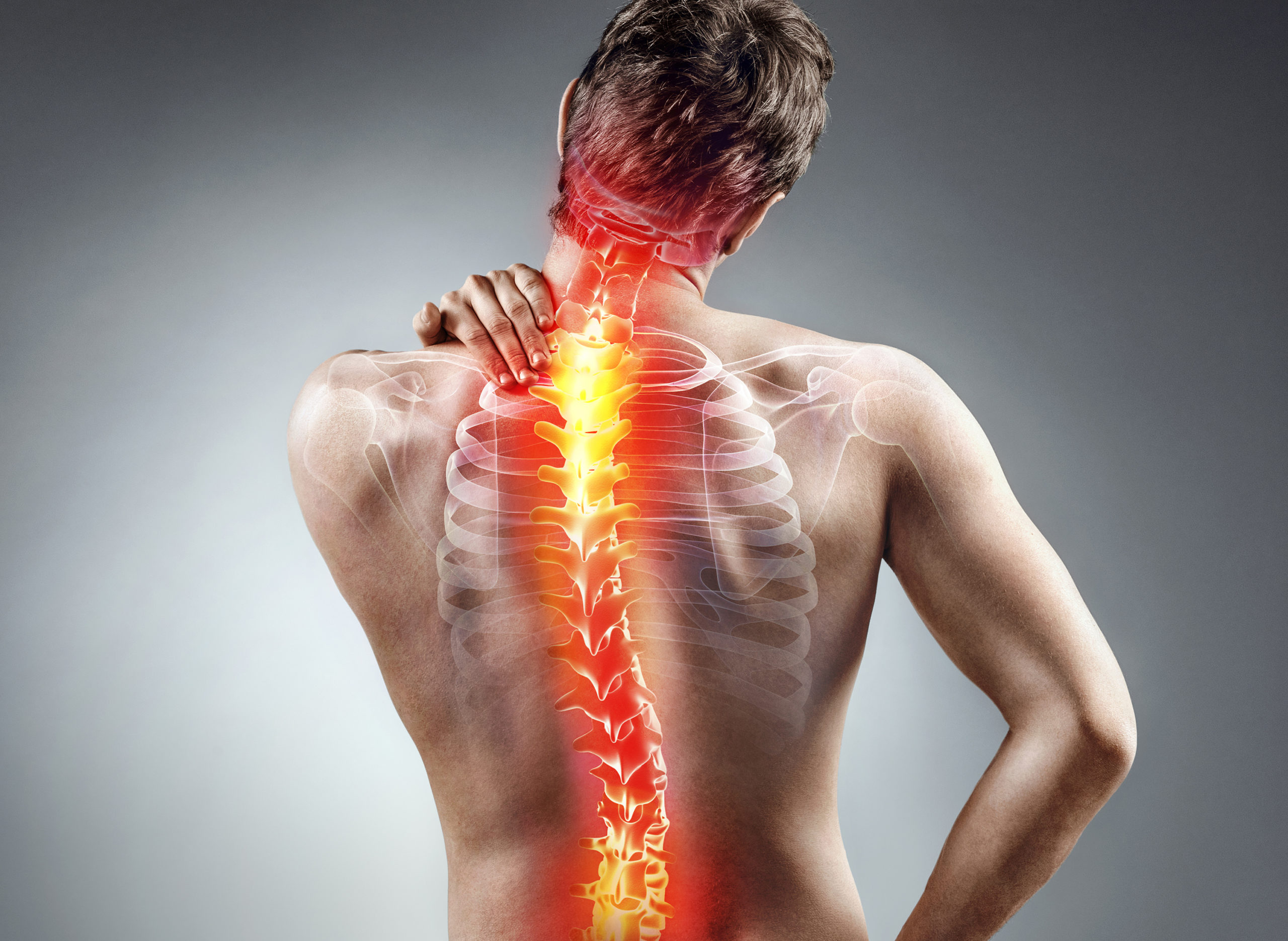
Back pain is a common ailment that affects millions of people worldwide. It can range from a dull, constant ache to a sudden, sharp pain that makes movement difficult. Understanding the anatomy of the back, the various causes of back pain, and the essential scans that might be needed for diagnosis can help in managing and treating this pervasive issue effectively.
Anatomy of the Back
The back is a complex structure made up of bones, muscles, ligaments, nerves, and other tissues. It can be divided into three main regions: the cervical (neck), thoracic (mid-back), and lumbar (lower back) spine.
1. Spinal Column
The spinal column, or backbone, is made up of 33 vertebrae stacked on top of each other. These vertebrae are divided into:
- Cervical vertebrae (C1-C7): These are the seven vertebrae in the neck.
- Thoracic vertebrae (T1-T12): These 12 vertebrae are located in the mid-back.
- Lumbar vertebrae (L1-L5): These five vertebrae are in the lower back and bear most of the body’s weight.
- Sacral vertebrae (S1-S5): These are fused vertebrae located below the lumbar region.
- Coccygeal vertebrae (tailbone): Usually four fused vertebrae at the very bottom of the spine.
2. Intervertebral Discs
Between each vertebra are intervertebral discs that act as cushions or shock absorbers. These discs have a tough outer layer called the annulus fibrosus and a soft, gel-like center called the nucleus pulposus.
3. Muscles and Ligaments
The back muscles and ligaments provide support and movement to the spine. Key muscle groups include the erector spinae, which help maintain an upright posture, and the multifidus muscles, which stabilize the spine.
4. Nerves
The spinal cord runs through the spinal column and branches out into spinal nerves that control muscle movements and transmit sensory information from the body to the brain.
Causes of Back Pain
Back pain can be caused by a variety of factors, ranging from acute injuries to chronic conditions. Understanding these causes is crucial for effective treatment.
1. Muscle or Ligament Strain
Heavy lifting, sudden movements, or poor posture can strain the muscles and ligaments in the back. These strains can cause acute pain and may lead to muscle spasms.
2. Herniated or Bulging Discs
The intervertebral discs can herniate or bulge, putting pressure on the spinal nerves. This can cause pain, numbness, or weakness in the back and legs.
3. Degenerative Disc Disease
With age, the intervertebral discs can wear down, leading to degenerative disc disease. This condition can cause chronic pain and reduced mobility.
4. Spinal Stenosis
Spinal stenosis is the narrowing of the spinal canal, which can compress the spinal cord and nerves. This condition is often caused by age-related changes and can lead to pain, numbness, and weakness.
5. Arthritis
Osteoarthritis can affect the spine, leading to the breakdown of cartilage between the vertebrae. This can result in pain and stiffness.
6. Skeletal Irregularities
Conditions such as scoliosis, which is an abnormal curvature of the spine, can cause back pain. Other skeletal irregularities like kyphosis and lordosis can also contribute to discomfort.
7. Osteoporosis
Osteoporosis is a condition where bones become brittle and fragile. Vertebral fractures due to osteoporosis can cause severe back pain.
8. Infections and Tumors
In rare cases, infections of the spine (such as osteomyelitis) or tumors can cause back pain. These conditions often require prompt medical attention.
Essential Scans for Diagnosing Back Pain
When back pain persists or is severe, medical imaging scans are often necessary to diagnose the underlying cause. Here are some essential scans that might be needed:
1. X-Rays
X-rays are often the first imaging test used to evaluate back pain. They can reveal bone abnormalities, fractures, and signs of arthritis. However, X-rays cannot show soft tissue structures like muscles, discs, or nerves.
2. Magnetic Resonance Imaging (MRI)
MRIs use powerful magnets and radio waves to create detailed images of the spine’s soft tissues, including the intervertebral discs, spinal cord, and nerves. MRIs are particularly useful for diagnosing herniated discs, spinal stenosis, and infections or tumors.
3. Computed Tomography (CT) Scans
CT scans use X-rays to create cross-sectional images of the spine. They provide more detailed information than regular X-rays and are useful for diagnosing spinal fractures, tumors, and spinal stenosis.
4. Bone Scans
Bone scans involve injecting a small amount of radioactive material into the bloodstream, which accumulates in areas of high bone activity. This scan can help detect bone infections, fractures, and tumors.
5. Electromyography (EMG)
EMG measures the electrical activity of muscles and nerves. It can help diagnose nerve compression or damage, such as that caused by a herniated disc or spinal stenosis.
Conclusion
Timely MRI scans, as provided by services like GetScanned, are a powerful tool for effectively managing and diagnosing back pain. By identifying the underlying causes of pain early, these scans can significantly improve treatment outcomes, reduce healthcare costs, and enhance patient empowerment. In the context of private versus NHS services, private MRI scans offer quicker access and more flexible scheduling, allowing for faster diagnosis and intervention than the often lengthy wait times associated with NHS scans. This advantage makes private MRI services an invaluable resource for those seeking prompt and efficient healthcare solutions for back pain.
Both patients and healthcare providers should consider the benefits of early MRI screening through private services like GetScanned as a proactive step towards better back pain management and overall well-being.

